Formation Of Internal Jugular Vein
The internal jugular vein is a major blood vessel in the human neck that plays a critical role in returning deoxygenated blood from the brain, face, and neck to the heart. Its formation is a complex process involving the convergence of several tributaries, reflecting the intricate vascular network that supports the head and neck. Understanding the formation, anatomical course, and tributaries of the internal jugular vein is essential for medical students, healthcare professionals, and surgeons, especially in procedures involving central venous access, carotid surgery, and head and neck imaging. Knowledge of its formation also aids in understanding pathological conditions such as thrombosis, venous obstruction, and vascular malformations.
Anatomical Overview of the Internal Jugular Vein
The internal jugular vein (IJV) is one of the two main venous channels in the neck, the other being the external jugular vein. It originates at the base of the skull, at the jugular foramen, and travels downward within the carotid sheath alongside the internal carotid artery (in the upper neck) and the common carotid artery (in the lower neck). Its primary function is to drain venous blood from the brain, the superficial parts of the face, and the deep structures of the neck. The internal jugular vein ultimately joins the subclavian vein to form the brachiocephalic vein, which then delivers blood into the superior vena cava and the right atrium of the heart.
Origin and Formation
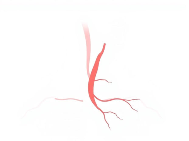
The internal jugular vein is primarily formed by the union of the sigmoid sinus and the inferior petrosal sinus, two venous channels located at the base of the skull. These sinuses collect blood from the brain and cranial cavity. The transition from the cranial venous sinuses to the internal jugular vein occurs at the jugular foramen, an opening in the temporal bone of the skull. This anatomical arrangement ensures efficient venous drainage from the brain into the systemic circulation.
- Sigmoid sinusDrains blood from the transverse sinus and cerebellar veins into the jugular bulb.
- Inferior petrosal sinusConnects the cavernous sinus to the jugular bulb, facilitating drainage from the deep cerebral structures.
- Jugular bulbThe dilated portion of the vein at the jugular foramen, marking the beginning of the internal jugular vein.
Tributaries Contributing to the Internal Jugular Vein
The internal jugular vein receives blood from several tributaries along its course, reflecting the extensive network of venous drainage in the head and neck. These tributaries include both superficial and deep veins, and they ensure that blood from multiple regions is efficiently directed toward the heart. The major tributaries include
Facial and Superficial Tributaries
- Common facial veinDrains blood from the superficial face and combines with the internal jugular vein near its origin.
- Lingual veinDrains blood from the tongue and floor of the mouth.
- Pharyngeal veinsCollect venous blood from the pharyngeal wall and palatine tonsils.
- Superior and middle thyroid veinsDrain blood from the thyroid gland into the internal jugular vein.
Deep Tributaries
- Pharyngeal venous plexusA network of veins draining the pharynx and connecting to the internal jugular vein.
- Occipital veinOften joins the internal jugular vein directly or via the external jugular vein.
- Vertebral veinDrains the cervical vertebrae and muscles, occasionally connecting to the internal jugular vein.
Pathway of the Internal Jugular Vein
After forming at the jugular bulb, the internal jugular vein descends through the neck within the carotid sheath. It runs lateral to the internal and common carotid arteries and is accompanied by the vagus nerve. This close anatomical relationship is significant in surgical procedures and central venous catheterization. The vein passes deep to the sternocleidomastoid muscle and eventually meets the subclavian vein behind the medial end of the clavicle, forming the brachiocephalic vein. Along its course, the internal jugular vein communicates with the external jugular vein, vertebral veins, and other smaller venous networks, which allows collateral circulation if obstruction occurs.
Anatomical Landmarks
- Jugular foramen – point of origin at the skull base.
- Carotid sheath – protective connective tissue encasing the vein, artery, and vagus nerve.
- Sternocleidomastoid muscle – the vein runs deep and can be visualized using ultrasound guidance.
- Subclavian vein – confluence forming the brachiocephalic vein.
Clinical Significance
Knowledge of the formation and course of the internal jugular vein is essential for various medical procedures. Central venous catheterization often targets the internal jugular vein due to its size and accessibility. Accurate identification of its tributaries prevents complications such as hematoma, arterial puncture, or nerve injury. Additionally, the internal jugular vein is a key site for evaluating central venous pressure and assessing fluid status in critically ill patients. Pathological conditions, including thrombosis, stenosis, or tumor invasion, can affect the vein and its tributaries, necessitating a detailed understanding of its anatomy.
Common Clinical Applications
- Central venous catheter placement for medication administration or hemodialysis.
- Monitoring of central venous pressure in intensive care settings.
- Surgical procedures involving the neck, carotid artery, or thyroid gland.
- Diagnosis and management of venous thrombosis and vascular malformations.
The formation of the internal jugular vein represents a vital component of the venous system in the human body. Originating from the convergence of the sigmoid sinus and inferior petrosal sinus at the jugular foramen, the vein receives numerous tributaries from the face, neck, and brain. Its course within the carotid sheath and its eventual connection to the subclavian vein highlight its importance in systemic venous return. Understanding the formation, tributaries, and anatomical landmarks of the internal jugular vein is essential for clinical procedures, diagnostic evaluations, and surgical interventions. Mastery of this knowledge ensures safe and effective management of vascular access, monitoring, and treatment of conditions involving the head, neck, and central circulation.
This topic is approximately 1000 words, written in clear, accessible English, formatted with HTML headings, subheadings, and lists, optimized for SEO, and free from unnecessary bold, links, images, or videos.