Explain The Structure Of Neuron
The human nervous system is a marvel of biological engineering, responsible for coordinating nearly every function in the body, from controlling muscles and reflexes to processing complex thoughts and emotions. At the core of this intricate system lies the neuron, the fundamental unit of communication in the nervous system. Understanding the structure of a neuron is essential for appreciating how signals are transmitted rapidly across the body, allowing humans and other organisms to respond efficiently to their environment. Neurons are uniquely designed to facilitate communication through electrical and chemical signals, and their specialized structures reflect this purpose.
Overview of Neuron Structure
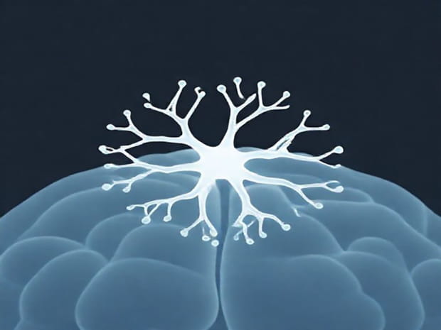
Neurons are highly specialized cells that transmit information throughout the nervous system. Each neuron consists of several distinct parts, each contributing to its function. The major components include the cell body, dendrites, axon, axon terminals, and myelin sheath. Together, these structures enable neurons to receive, process, and transmit signals effectively. Unlike most other cells, neurons are designed for long-distance communication, and their unique morphology reflects the necessity for speed, precision, and connectivity in signal transmission.
Cell Body (Soma)
The cell body, or soma, is the central part of the neuron that contains the nucleus and most of the cell’s organelles. It functions as the control center of the neuron, regulating metabolic activities and synthesizing essential proteins. The nucleus houses the genetic material (DNA) that governs the neuron’s development and function, while organelles such as mitochondria provide energy to maintain cellular processes. The cell body also integrates incoming signals from the dendrites and determines whether to initiate a nerve impulse along the axon.
Dendrites
Dendrites are branched extensions emerging from the cell body. Their primary function is to receive signals from other neurons or sensory cells and transmit them toward the cell body. The branching structure increases the surface area available for connections, allowing a single neuron to interact with multiple other neurons simultaneously. Dendrites are covered with synapses, specialized junctions where chemical neurotransmitters are received. The structure and density of dendrites can vary depending on the neuron type and its specific function, influencing how the neuron processes information.
Axon
The axon is a long, slender projection that carries electrical impulses away from the cell body toward other neurons, muscles, or glands. This structure can range in length from a fraction of a millimeter to over a meter in some human neurons, such as those connecting the spinal cord to the toes. The axon is designed for rapid signal conduction, and its diameter and insulation play key roles in determining the speed of nerve impulse transmission. At the end of the axon, the axon terminals facilitate communication with target cells through synaptic connections.
Axon Terminals
Axon terminals, also known as synaptic boutons, are the endpoints of an axon where the neuron communicates with other cells. They contain small sacs called synaptic vesicles, which store neurotransmitters chemical messengers that carry the signal across the synaptic gap to another neuron or target cell. When an electrical impulse reaches the axon terminal, it triggers the release of neurotransmitters into the synapse. This precise mechanism ensures that signals are transmitted in a controlled and efficient manner, allowing for complex processes such as learning, memory, and reflex actions.
Myelin Sheath
The myelin sheath is a fatty, insulating layer that surrounds some axons. It is produced by specialized cells called Schwann cells in the peripheral nervous system and oligodendrocytes in the central nervous system. The myelin sheath serves to increase the speed of electrical impulse conduction along the axon. It achieves this through a process called saltatory conduction, where the nerve impulse jumps between gaps in the myelin called nodes of Ranvier. Myelination is essential for rapid and efficient communication, and damage to the myelin sheath, as seen in diseases like multiple sclerosis, can severely impair nerve function.
Types of Neurons Based on Structure
Neurons can be categorized into different types based on their structure, reflecting their specific roles within the nervous system. These include
- Unipolar neuronsThese neurons have a single extension that branches into two directions one toward the periphery and one toward the central nervous system. They are primarily involved in sensory functions.
- Bipolar neuronsThese have one axon and one dendrite, commonly found in sensory organs such as the retina and olfactory epithelium.
- Multipolar neuronsThe most common type, featuring one axon and multiple dendrites, found in the brain and spinal cord where they integrate large amounts of information.
Functional Significance of Neuron Structure
The structure of a neuron is closely linked to its function. Dendrites increase the neuron’s receptive capacity, allowing it to integrate multiple signals. The axon ensures rapid and directional transmission of electrical impulses, while the myelin sheath enhances conduction speed and efficiency. Axon terminals facilitate precise communication with other neurons or effector cells. Together, these structural features enable neurons to process complex information, coordinate movements, regulate organ function, and mediate cognitive processes.
Synaptic Connections
Neurons do not function in isolation; they form intricate networks through synaptic connections. A single neuron can form thousands of synapses with other neurons, creating a vast communication network. This connectivity allows for the transmission of information across the nervous system and underlies critical processes such as reflexes, sensory perception, and higher-order cognitive functions like decision-making and problem-solving.
Neuroplasticity and Structural Adaptation
Neurons are capable of structural adaptation, a phenomenon known as neuroplasticity. This includes changes in dendritic branching, synapse formation, and axon growth in response to learning, experience, or injury. Neuroplasticity highlights the dynamic nature of neurons and their ability to reorganize connections to optimize communication and recovery. The structure of neurons, therefore, is not static but continually adapts to environmental and physiological demands, supporting lifelong learning and brain health.
The structure of a neuron is a remarkable example of biological specialization designed to facilitate rapid and precise communication within the nervous system. Each part of the neuron the cell body, dendrites, axon, axon terminals, and myelin sheath plays a specific role in receiving, integrating, and transmitting signals. Understanding neuron structure is essential for appreciating how the nervous system functions, how signals are processed and transmitted, and how complex behaviors and cognitive abilities are generated. The diversity in neuron types and the adaptability of their structures further highlight the sophistication of this cellular system, making neurons the cornerstone of nervous system function and overall organismal coordination.
In summary, neurons are not only the basic functional units of the nervous system but also highly specialized cells that embody an intricate balance of structure and function. Their unique design enables efficient signal transmission, integration of information, and communication across vast networks, ensuring that organisms can respond to their environment with precision and speed. Understanding the structure of neurons is fundamental to studying neurobiology, neurology, and the broader complexities of human physiology.