Draw The Structure Of Neuron
Neurons are the fundamental units of the nervous system, responsible for transmitting information throughout the body. They are specialized cells that communicate via electrical and chemical signals, allowing us to sense the environment, control muscles, and process thoughts. Understanding the structure of a neuron is crucial for studying how the nervous system functions. Each neuron has unique components that work together to receive, process, and transmit signals efficiently. While drawing a neuron can help visualize its complex structure, comprehending each part’s role is equally important for students and researchers in neuroscience.
Overview of Neuron Structure
A neuron consists of several key components the cell body (soma), dendrites, axon, myelin sheath, axon terminals, and synaptic terminals. Each part has a specific function in the process of neural communication. The cell body contains the nucleus and organelles necessary for cell maintenance, while dendrites receive incoming signals from other neurons. The axon carries electrical impulses away from the cell body, often covered by a myelin sheath to increase conduction speed. At the end of the axon, axon terminals and synaptic terminals transmit signals to adjacent neurons or target cells.
Cell Body (Soma)
The cell body, or soma, is the central part of the neuron. It contains the nucleus, which houses genetic material and regulates cellular activities. Organelles such as mitochondria, endoplasmic reticulum, and Golgi apparatus support energy production, protein synthesis, and cellular maintenance. The soma integrates signals received from dendrites and initiates the action potential if the threshold is reached. Drawing the soma is typically represented as a rounded or oval shape with a prominent nucleus inside.
Dendrites
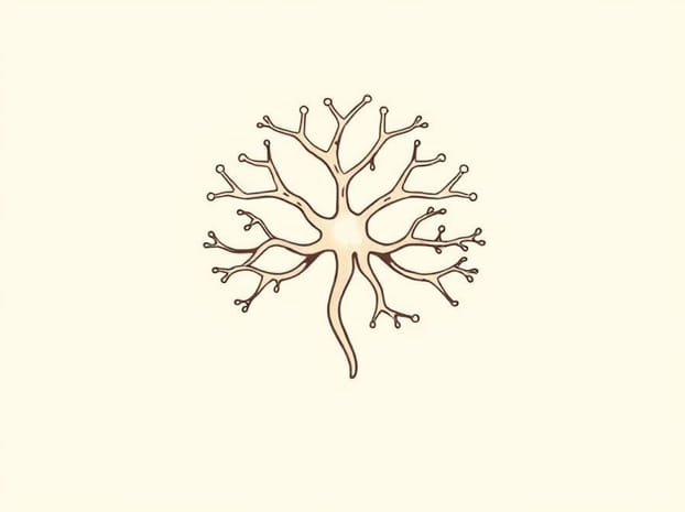
Dendrites are branch-like extensions from the soma. They receive electrical and chemical signals from other neurons’ axon terminals. Dendritic spines increase the surface area for synaptic connections, allowing neurons to form complex networks. In a neuron drawing, dendrites are depicted as multiple thin, tree-like branches radiating from the cell body. Their intricate design emphasizes their role in signal reception and integration.
Axon
The axon is a long, slender projection that transmits electrical impulses from the soma to other neurons or target cells. The length of the axon can vary from a few millimeters to over a meter, depending on the type of neuron. Axons often branch at their ends to form axon terminals, facilitating communication with multiple cells. In a neuron drawing, the axon is usually shown as a single elongated line extending from the soma, highlighting its role in signal propagation.
Myelin Sheath and Nodes of Ranvier
Many axons are wrapped in a myelin sheath, composed of lipid-rich Schwann cells or oligodendrocytes. This sheath insulates the axon and allows rapid transmission of electrical impulses through saltatory conduction. The nodes of Ranvier are small gaps between myelin segments where ion exchange occurs, enabling the action potential to jump along the axon efficiently. When illustrating a neuron, the myelin sheath can be represented as segmented coverings along the axon, with small interruptions to indicate nodes of Ranvier.
Axon Terminals and Synaptic Terminals
At the end of the axon, axon terminals (also called synaptic terminals) form connections with target cells, including other neurons, muscles, or glands. These terminals contain synaptic vesicles filled with neurotransmitters, which are released into the synaptic cleft to transmit signals chemically. In drawings, axon terminals are shown as small bulbous structures at the ends of axon branches, emphasizing their role in communication between cells.
Types of Neurons and Structural Variations
Neurons can be classified based on their structure and function. Multipolar neurons, which have multiple dendrites and one axon, are the most common and typically found in the central nervous system. Bipolar neurons have one dendrite and one axon and are usually involved in sensory pathways, such as in the retina. Unipolar neurons have a single projection that splits into two branches, one connecting to sensory receptors and the other to the spinal cord. Drawing different types of neurons involves adjusting the number and arrangement of dendrites and axons to reflect these structural variations.
Functional Aspects of Neuron Structure
The structure of a neuron is closely tied to its function. Dendrites allow extensive input integration, while the axon ensures that signals are transmitted rapidly over long distances. Myelin sheaths increase conduction velocity, which is critical for reflexes and coordinated movements. Axon terminals enable precise communication with target cells through synaptic transmission. Understanding these functional aspects helps in creating accurate and meaningful neuron diagrams that convey both structure and purpose.
Tips for Drawing a Neuron
- Start with the soma Draw a central oval or round shape to represent the cell body.
- Add the nucleus Inside the soma, draw a smaller circle for the nucleus.
- Include dendrites Extend multiple branched lines from the soma to represent dendrites.
- Draw the axon Extend a long line from the soma to indicate the axon’s length.
- Add myelin sheath Represent the myelin as segmented coverings along the axon, leaving gaps for nodes of Ranvier.
- Include axon terminals Draw small bulbous endings at the axon’s branches to show synaptic terminals.
- Label parts Clearly label soma, dendrites, axon, myelin sheath, nodes of Ranvier, and axon terminals for clarity.
Importance of Neuron Diagrams in Education
Drawing neuron structures is a valuable educational tool. Visual representations help students understand the complex architecture of neurons and their functional roles. Diagrams also facilitate learning about neural circuits, synaptic transmission, and the differences between neuron types. Teachers and researchers often use neuron illustrations to explain neurological processes, making them an essential component of neuroscience education and communication.
Understanding the structure of a neuron is fundamental for exploring how the nervous system functions. Each part of the neuron, from dendrites to axon terminals, plays a critical role in receiving, processing, and transmitting signals. Drawing a neuron not only helps visualize these components but also reinforces the functional relationships between them. By combining accurate illustrations with explanations of each structure’s role, learners can gain a comprehensive understanding of neuronal function, paving the way for further studies in neurobiology, neuroscience, and medical research.