Diagram Of Tsetse Fly
The tsetse fly is a medically significant insect native to sub-Saharan Africa, primarily known for its role in transmitting trypanosomiasis, commonly called sleeping sickness in humans and nagana in animals. Understanding the anatomy and structure of the tsetse fly is essential for entomologists, epidemiologists, and public health professionals. A diagram of the tsetse fly helps visualize its distinct body parts, feeding mechanisms, and adaptations that enable it to thrive in various ecological niches. Studying the tsetse fly’s morphology provides insights into its life cycle, behavior, and strategies for disease control. This topic explores the key anatomical features of the tsetse fly, their functions, and how a detailed diagram can enhance comprehension of this important vector species.
Overview of the Tsetse Fly
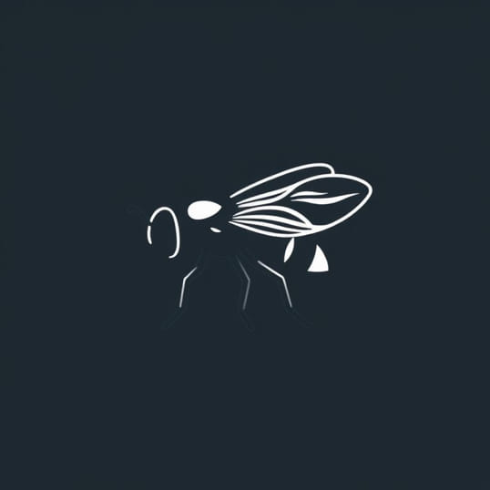
Tsetse flies belong to the genusGlossinaand are hematophagous insects, meaning they feed exclusively on the blood of vertebrate hosts. They are medium-sized flies with a characteristic wing structure and forward-pointing proboscis adapted for piercing skin and sucking blood. Tsetse flies are highly specialized vectors of trypanosomes, protozoan parasites responsible for sleeping sickness. Their unique anatomical adaptations, captured in scientific diagrams, illustrate the features that facilitate blood feeding, reproduction, and disease transmission. A visual representation, such as a diagram, helps clarify complex structures that are difficult to describe textually.
External Anatomy
The external anatomy of the tsetse fly includes several key components that are essential for survival and function. A diagram typically highlights these features in a labeled format, allowing for easy identification.
- HeadThe head houses sensory organs such as the compound eyes, antennae, and mouthparts. The compound eyes provide a wide field of vision, while the antennae detect chemical cues from hosts. The proboscis is a tubular feeding structure used to pierce skin and extract blood.
- ThoraxThe thorax is the central body segment where wings and legs attach. Muscles within the thorax facilitate flight and movement. Diagrams often show the arrangement of wings and halteres, which help maintain stability during flight.
- AbdomenThe abdomen contains digestive and reproductive organs. In female tsetse flies, the abdomen houses specialized structures for viviparous reproduction, where larvae develop internally before being deposited as pupae.
- LegsTsetse flies have six jointed legs adapted for walking and clinging to hosts. The tarsal segments often end in claws or pads, aiding in grip on animal skin or vegetation.
Mouthparts and Feeding Mechanism
The tsetse fly’s mouthparts are uniquely adapted for hematophagy and are prominently displayed in anatomical diagrams. The proboscis is elongated, rigid, and reinforced with a labrum and hypopharynx that work together to penetrate host skin and locate blood vessels. Salivary glands inject anticoagulant compounds, which prevent blood clotting and facilitate smooth feeding. Understanding the structure of the proboscis is crucial in studying how trypanosomes are transmitted from host to host during blood meals.
Functional Anatomy of the Proboscis
- LabrumActs as a protective sheath for other mouthparts and assists in guiding the proboscis into host tissue.
- HypopharynxDelivers saliva containing anticoagulants and trypanosomes into the host during feeding.
- Mandibles and MaxillaeHelp pierce skin and facilitate the insertion of the proboscis.
Reproductive System
Tsetse flies exhibit unique reproductive strategies, including viviparity, which is rare among insects. A diagram of the tsetse fly often illustrates the female reproductive system, including the uterus and specialized glands that nourish the developing larva. Females produce a single larva at a time, which is retained internally and nourished with milk-like secretions until it is ready to pupate. This adaptation ensures higher survival rates for offspring compared to egg-laying insects.
Male Reproductive Anatomy
The male reproductive system, illustrated in detailed diagrams, includes testes, accessory glands, and aedeagus. These structures are involved in sperm production, transfer, and mating behavior. Understanding the male reproductive anatomy is essential for studies on tsetse fly population control and sterilization techniques, which are part of integrated vector management programs.
Internal Digestive System
The tsetse fly’s digestive system is specialized for processing blood meals. Diagrams typically label the foregut, midgut, and hindgut. The midgut houses symbiotic bacteria that assist in nutrient absorption and trypanosome development. The digestive system also includes salivary glands connected to the proboscis, which play a critical role in both feeding and pathogen transmission.
Role in Disease Transmission
The anatomy of the digestive and salivary systems is central to the tsetse fly’s ability to transmit trypanosomes. Blood ingested by the fly is first processed in the midgut, where parasites multiply before migrating to the salivary glands. A well-labeled diagram can clearly show this pathway, making it easier for researchers to understand infection dynamics and develop control strategies.
Importance of Diagrams in Understanding Tsetse Fly Anatomy
Visual representations, such as diagrams, are indispensable for studying the tsetse fly. They provide a clear overview of external and internal anatomy, allow identification of critical functional structures, and help researchers, students, and health workers understand the complex relationships between anatomy and disease transmission. Diagrams are also used in educational materials to illustrate differences between tsetse fly species, their feeding mechanisms, and life cycle stages.
Educational and Research Applications
- Vector Biology StudiesDiagrams help identify anatomical features related to pathogen transmission.
- Public Health TrainingVisual aids assist health workers in understanding how tsetse flies spread disease.
- Taxonomic IdentificationDetailed illustrations highlight distinguishing characteristics used to differentiate species.
- Control StrategiesUnderstanding anatomy supports the design of traps, insecticides, and sterilization programs.
The tsetse fly is a remarkable insect with unique adaptations for blood feeding, reproduction, and survival in its natural environment. A diagram of the tsetse fly provides a comprehensive view of its external and internal anatomy, highlighting features such as the proboscis, compound eyes, wings, digestive system, and reproductive organs. These visual representations are essential for understanding its role as a disease vector, guiding research, and developing effective control strategies. By studying tsetse fly anatomy through diagrams, entomologists and public health professionals can gain critical insights into its biology, behavior, and impact on human and animal health. From feeding mechanisms to reproductive strategies, the tsetse fly serves as a key model for understanding vector biology and the challenges associated with managing vector-borne diseases in Africa.