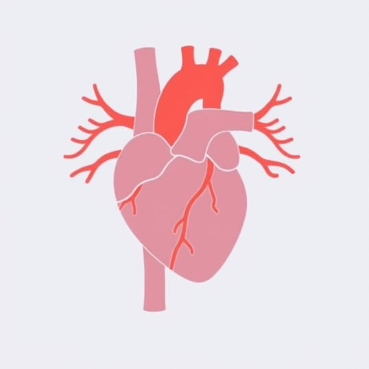
Diagram Of Coronary Arteries
Understanding the diagram of coronary arteries is an important step in learning how the heart functions and how blood reaches its vital tissues. The coronary arteries are responsible for delivering oxygen-rich blood to the heart muscle itself, ensuring that it continues to pump efficiently. Without this supply, the heart would not be able to sustain life. Medical students, healthcare professionals, and even patients are often introduced to a coronary artery diagram to visualize how these vessels branch across the heart. Knowing where the left and right coronary arteries, as well as their branches, are located helps in diagnosing and treating conditions such as coronary artery disease, heart attacks, and angina.
Basic Structure of Coronary Arteries
Coronary arteries are specialized blood vessels that sit on the surface of the heart. They emerge from the base of the aorta, the main artery that carries blood from the heart to the rest of the body. In most diagrams of coronary arteries, two main vessels are shown the left coronary artery (LCA) and the right coronary artery (RCA). Each of these branches further into smaller arteries that supply specific regions of the heart muscle.
Left Coronary Artery (LCA)
The left coronary artery plays a major role in supplying blood to the left side of the heart, which is responsible for pumping oxygenated blood throughout the body. The LCA typically divides into two important branches
- Left Anterior Descending (LAD) ArterySupplies the front wall of the left ventricle and the interventricular septum.
- Left Circumflex (LCx) ArteryWraps around the heart and delivers blood to the left atrium and the side and back of the left ventricle.
Right Coronary Artery (RCA)
The right coronary artery mainly supplies the right side of the heart, including the right atrium, right ventricle, and parts of the conduction system that regulate heartbeat. The RCA gives off important branches such as
- Right Marginal ArterySupplies the right ventricular wall.
- Posterior Descending Artery (PDA)Supplies the back portion of the interventricular septum and part of the ventricles.
Coronary Artery Diagram and Its Importance
A diagram of coronary arteries serves as a visual guide to understand the layout and function of these vessels. It shows the pathways of the arteries and how they wrap around the heart. This kind of diagram is not only useful for educational purposes but also for clinical practice. Cardiologists often refer to it when explaining to patients where blockages occur and how procedures like angioplasty or bypass surgery will help.
Educational Use
For students, the diagram highlights how the left and right arteries divide and distribute blood. It provides a roadmap for understanding how oxygen is delivered to the heart muscle. By studying the diagram, learners can connect the structure of the heart with its function.
Clinical Use
In hospitals, a coronary artery diagram is often shown to patients undergoing cardiac procedures. It simplifies explanations and helps patients understand which part of the heart is affected. Surgeons also use such diagrams to plan bypass surgeries by mapping where new vessels will be grafted.
Branches of Coronary Arteries in Detail
Beyond the primary arteries, the coronary circulation involves a detailed network of smaller branches. These vessels ensure that all areas of the heart muscle receive adequate blood supply. A comprehensive diagram of coronary arteries usually depicts the following
- Diagonal BranchesBranch from the LAD and supply the front of the left ventricle.
- Obtuse Marginal BranchesExtend from the LCx and supply the side of the left ventricle.
- Sinoatrial Nodal ArteryOften arises from the RCA and supplies the SA node, the heart’s natural pacemaker.
- Atrioventricular Nodal ArterySupplies the AV node, crucial for coordinating heartbeat.
Blood Flow and Function
The diagram of coronary arteries also illustrates how blood flows from the aorta into these vessels. Blood rich in oxygen and nutrients enters the coronary arteries during diastole, when the heart muscle is relaxed. This ensures that the heart muscle receives its blood supply without interruption. Each artery is responsible for delivering blood to specific regions, and any blockage can have serious consequences depending on the location.
Coronary Artery Disease
When plaques or fatty deposits block these vessels, coronary artery disease develops. The diagram is crucial in identifying the exact site of narrowing. For example, a blockage in the LAD artery is sometimes called the widow-maker because it can be life-threatening due to the large portion of the heart it supplies.
Variations in Coronary Anatomy
Not all coronary artery diagrams look identical because human anatomy can vary. Some individuals may have dominant circulation through the RCA, while others rely more on the LCx. A detailed diagram often indicates these variations so that doctors can plan treatments effectively.
Modern Imaging and Diagrams
While traditional diagrams are drawn schematically, modern technology provides even more accurate views of coronary arteries. CT angiography and 3D imaging produce detailed diagrams showing patient-specific coronary anatomy. These advances make diagnosis and treatment more precise.
3D Reconstruction
With the use of advanced imaging, diagrams can now be created based on real patient data. This allows cardiologists to visualize the exact location of blockages, plan stent placement, and monitor outcomes after surgery.
Practical Applications of Coronary Artery Diagrams
Beyond medical studies, the diagram of coronary arteries has a role in fitness, health awareness, and patient education. It helps the general public understand why lifestyle choices like diet, exercise, and avoiding smoking are important for heart health. By connecting these choices to the actual pathways of blood supply, diagrams make the information tangible and easy to grasp.
The diagram of coronary arteries is more than just a picture it is a fundamental tool in understanding how the heart functions and how blood is distributed to keep it working. By visualizing the left and right coronary arteries and their branches, people gain a clear understanding of how vital these vessels are. From medical students to heart patients, everyone can benefit from studying such diagrams. They highlight the importance of maintaining healthy arteries and provide essential guidance for diagnosing and treating cardiovascular diseases. With modern imaging techniques, these diagrams continue to evolve, giving us clearer insights into the heart’s lifeline.