Evaluation Of Cerebellar Ataxic Patients
Cerebellar ataxia is a neurological condition that affects coordination, balance, and motor control, often resulting from damage or dysfunction in the cerebellum. Patients with cerebellar ataxia may present with a range of symptoms, including unsteady gait, difficulty with fine motor tasks, tremors, and impaired speech. Evaluating these patients requires a systematic and thorough approach to determine the underlying cause, assess the severity of the condition, and guide treatment planning. A comprehensive evaluation not only helps in accurate diagnosis but also informs interventions that can improve quality of life and functional independence for affected individuals. Understanding the nuances of cerebellar ataxia evaluation is essential for neurologists, rehabilitation specialists, and allied healthcare professionals.
Clinical History and Symptom Assessment
The evaluation of cerebellar ataxic patients begins with a detailed clinical history. Healthcare providers need to inquire about the onset, duration, and progression of symptoms. Sudden onset may suggest a vascular event such as a cerebellar stroke, whereas gradual progression could indicate degenerative diseases like spinocerebellar ataxia. It is crucial to document family history, as many cerebellar disorders have a genetic component. Additionally, understanding associated symptoms, such as dizziness, nausea, or cognitive changes, provides clues about the extent of cerebellar involvement and potential comorbid conditions.
Key Historical Elements
- Onset and progression of coordination problems.
- Family history of neurological or genetic disorders.
- Presence of associated neurological symptoms like tremors or dysarthria.
- Exposure to toxins, medications, or infections that may affect cerebellar function.
- Impact on daily activities and functional limitations.
Physical Examination
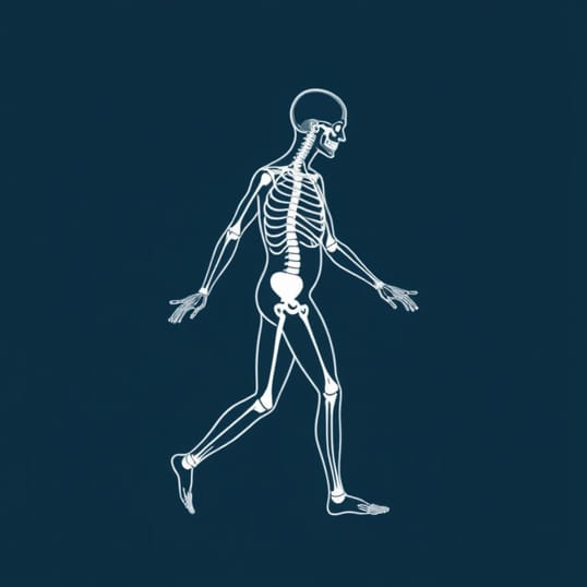
The physical examination of cerebellar ataxic patients focuses on identifying signs of cerebellar dysfunction. Standardized neurological examinations can reveal abnormalities in gait, posture, and coordination. Observing the patient’s walking pattern can indicate truncal instability or wide-based gait, which are characteristic of cerebellar involvement. Testing fine motor skills, such as finger-to-nose or heel-to-shin movements, helps identify dysmetria. Additionally, assessing speech, eye movements, and reflexes provides further insight into the cerebellar pathology.
Examination Techniques
- Gait assessment Observing for unsteady, broad-based walking patterns.
- Coordination tests Finger-to-nose, rapid alternating movements, heel-to-shin testing.
- Speech evaluation Checking for slurred or scanning speech (dysarthria).
- Oculomotor assessment Evaluating for nystagmus, abnormal saccades, or impaired smooth pursuit.
- Muscle tone and reflexes Assessing hypotonia and changes in deep tendon reflexes.
Laboratory Investigations
Laboratory tests play a critical role in evaluating cerebellar ataxic patients, particularly to identify reversible causes. Blood tests may include assessments of vitamin levels (such as vitamin B12 and E), thyroid function, liver and kidney function, and markers of autoimmune disease. Screening for metabolic or toxic causes, including heavy metal exposure or chronic alcohol use, is also important. These investigations can help differentiate between acquired ataxias and hereditary or degenerative forms.
Common Laboratory Tests
- Complete blood count and metabolic panel.
- Vitamin B12, vitamin E, and folate levels.
- Thyroid function tests.
- Autoimmune markers (e.g., anti-GAD antibodies).
- Toxin screening, including alcohol and heavy metals.
Neuroimaging
Neuroimaging is essential for visualizing structural or functional abnormalities in the cerebellum. Magnetic Resonance Imaging (MRI) is the preferred modality, providing detailed views of cerebellar anatomy and detecting lesions, atrophy, or tumors. Computed Tomography (CT) scans may be used in acute settings, such as suspected stroke, to rapidly identify hemorrhage or infarction. Advanced imaging techniques, including diffusion tensor imaging or functional MRI, can provide additional information about cerebellar connectivity and function in research or complex clinical cases.
Imaging Considerations
- MRI for structural assessment Detects atrophy, tumors, or demyelinating lesions.
- CT scan for acute events Rapid identification of hemorrhage or infarction.
- Functional imaging Evaluates cerebellar activity and neural networks.
- Serial imaging Monitors disease progression in degenerative ataxias.
Electrophysiological Studies
Electrophysiological studies, such as electroencephalography (EEG) and electromyography (EMG), can be valuable adjuncts in the evaluation of cerebellar ataxia. While EEG primarily assesses cortical activity, it may help rule out seizure-related causes of motor disturbances. EMG and nerve conduction studies are particularly useful for differentiating cerebellar ataxia from peripheral neuropathies, which can present with similar balance or coordination issues. These studies contribute to a comprehensive understanding of the neuromuscular system and help refine the diagnosis.
Utility of Electrophysiological Studies
- EMG Differentiates peripheral neuropathy from cerebellar pathology.
- Nerve conduction studies Evaluates sensory and motor nerve integrity.
- EEG Assesses cortical involvement and rules out seizure disorders.
- Evoked potentials Can detect subclinical cerebellar or sensory pathway dysfunction.
Genetic and Molecular Testing
For patients with suspected hereditary ataxia, genetic testing is a key component of the evaluation. Testing can identify specific gene mutations associated with various spinocerebellar ataxias, Friedreich’s ataxia, or episodic ataxias. Molecular testing aids in confirming the diagnosis, providing prognostic information, and informing family counseling. In some cases, identifying a genetic cause can also guide targeted therapies or participation in clinical trials.
Approach to Genetic Testing
- Family history analysis to identify inheritance patterns.
- Targeted gene panels for common ataxia-related genes.
- Whole exome or genome sequencing for undiagnosed cases.
- Genetic counseling for patients and family members.
Functional Assessment
Assessing functional abilities is an important part of evaluating cerebellar ataxic patients. Standardized tools, such as the Scale for the Assessment and Rating of Ataxia (SARA) or the International Cooperative Ataxia Rating Scale (ICARS), provide objective measures of motor function, balance, and coordination. Functional assessment helps track disease progression, evaluate treatment efficacy, and develop personalized rehabilitation plans. Occupational and physical therapy evaluations can further assess the impact on daily living activities and guide interventions to enhance independence.
Functional Assessment Tools
- Scale for the Assessment and Rating of Ataxia (SARA).
- International Cooperative Ataxia Rating Scale (ICARS).
- Timed up-and-go test for mobility assessment.
- Balance and gait analysis.
- Fine motor skill evaluations.
Evaluating cerebellar ataxic patients requires a comprehensive, multidisciplinary approach that integrates clinical history, physical examination, laboratory investigations, neuroimaging, electrophysiological studies, genetic testing, and functional assessments. Each component provides crucial information that contributes to accurate diagnosis, prognosis, and individualized management strategies. Timely and thorough evaluation is essential not only for addressing the underlying cause but also for optimizing patient outcomes through targeted interventions, rehabilitation, and supportive care. By understanding the full spectrum of evaluation techniques, healthcare professionals can provide effective and compassionate care for individuals living with cerebellar ataxia.
Summary of Evaluation Steps
- Detailed clinical history to understand symptom onset, progression, and family history.
- Comprehensive physical and neurological examination, including gait, coordination, speech, and eye movements.
- Laboratory investigations to identify reversible metabolic, toxic, or autoimmune causes.
- Neuroimaging with MRI or CT to detect structural abnormalities or acute events.
- Electrophysiological studies to differentiate peripheral neuropathies and assess neuromuscular function.
- Genetic testing for hereditary ataxias and molecular diagnosis.
- Functional assessment using standardized rating scales and therapy evaluations.