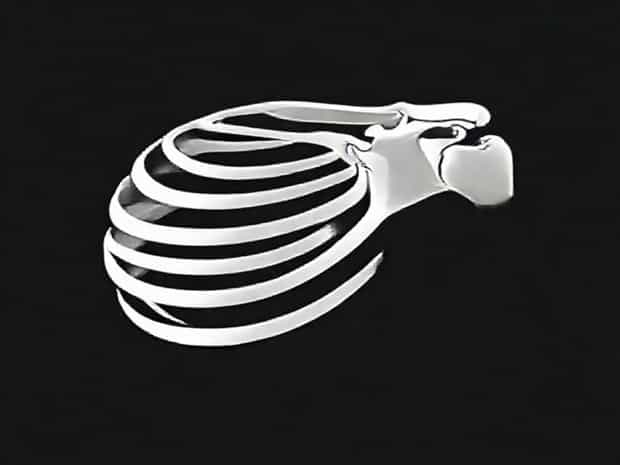
Intertubercular Sulcus Of Humerus
The intertubercular sulcus of the humerus, also known as the bicipital groove, is an important anatomical feature of the upper arm bone that plays a crucial role in shoulder movement and muscle attachment. This groove is located on the anterior surface of the humerus, running between the greater and lesser tubercles, and serves as a channel for the tendon of the long head of the biceps brachii muscle. Understanding the anatomy, function, and clinical significance of the intertubercular sulcus is essential for medical students, orthopedic specialists, and anyone interested in musculoskeletal health and shoulder mechanics.
Anatomical Features of the Intertubercular Sulcus
The intertubercular sulcus is a longitudinal groove situated on the proximal end of the humerus. It extends downward from the lesser tubercle medially to the greater tubercle laterally. The walls of the sulcus serve as attachment sites for various muscles and ligaments, providing stability and facilitating coordinated movement of the shoulder joint. The floor of the groove is often roughened and serves as an insertion point for the latissimus dorsi tendon. Its unique shape allows for the smooth passage of the biceps tendon, preventing friction and facilitating efficient movement.
Relationships with Surrounding Structures
The intertubercular sulcus is intimately associated with several key anatomical structures in the shoulder region
- Greater tubercleLateral border of the sulcus, serving as an attachment for the supraspinatus, infraspinatus, and teres minor muscles.
- Lesser tubercleMedial border, providing insertion for the subscapularis muscle.
- Biceps brachii tendonThe long head of the biceps tendon passes through the groove, held in place by the transverse humeral ligament.
- Transverse humeral ligamentSpans the sulcus to stabilize the biceps tendon and prevent dislocation.
- Latissimus dorsi insertionInserts at the floor of the sulcus, contributing to internal rotation and adduction of the arm.
These relationships highlight the sulcus’s role as a central hub for muscular attachment and tendon passage, essential for upper limb mobility.
Functional Significance
The intertubercular sulcus of the humerus is vital for the proper functioning of the shoulder joint. It serves primarily as a passageway for the long head of the biceps brachii tendon, allowing smooth gliding during arm flexion, abduction, and rotation. The biceps brachii muscle contributes to elbow flexion, forearm supination, and stabilization of the humeral head within the glenoid cavity. By guiding the tendon along the humerus, the sulcus prevents mechanical stress and potential injury, ensuring efficient movement and force transmission.
Role in Muscle Attachment
Besides guiding the biceps tendon, the intertubercular sulcus provides attachment points for major muscles that control arm movements. The latissimus dorsi attaches at the sulcus floor, aiding in internal rotation, adduction, and extension of the humerus. The pectoralis major inserts along the lateral lip of the sulcus, playing a key role in shoulder flexion, adduction, and internal rotation. These muscular connections make the sulcus integral to upper limb biomechanics, influencing posture, strength, and range of motion.
Clinical Relevance
Understanding the intertubercular sulcus is crucial in clinical practice, particularly in orthopedics and sports medicine. Injuries involving the long head of the biceps tendon, such as tendinitis or tears, often affect this groove. Overuse, repetitive overhead activities, or trauma can lead to inflammation of the tendon, causing pain localized to the anterior shoulder. Imaging studies, such as MRI or ultrasound, often focus on the sulcus to assess tendon integrity and guide treatment decisions.
Bicipital Tendon Disorders
The bicipital groove can be a site of tendon instability or dislocation. When the transverse humeral ligament is compromised, the tendon may slip out of the sulcus, resulting in discomfort, snapping, or limited arm function. Rehabilitation exercises, anti-inflammatory treatments, or surgical interventions may be necessary depending on severity. Knowledge of the sulcus anatomy aids surgeons in procedures like biceps tenodesis or tendon repair, ensuring proper alignment and restoration of function.
Implications for Shoulder Surgery
The intertubercular sulcus is often considered during shoulder arthroscopy, rotator cuff repair, and prosthetic implant placement. Accurate identification of the sulcus and surrounding landmarks is essential to avoid tendon damage and optimize post-surgical outcomes. Surgeons rely on a clear understanding of the sulcus anatomy to navigate complex procedures, prevent complications, and ensure effective rehabilitation.
Variations and Anatomical Differences
Anatomical variations of the intertubercular sulcus may occur between individuals. The depth, width, and curvature of the groove can differ, influencing biceps tendon stability and susceptibility to injury. Some individuals have a shallower sulcus, increasing the risk of tendon subluxation, while a deeper groove may provide additional stabilization. Recognizing these variations is important for personalized medical care, surgical planning, and tailored physical therapy approaches.
Imaging and Diagnostic Techniques
Modern imaging techniques allow detailed visualization of the intertubercular sulcus and its contents. MRI provides high-resolution images of the biceps tendon, surrounding ligaments, and muscle attachments. Ultrasound imaging is a non-invasive method to assess dynamic tendon movement within the sulcus, useful for diagnosing instability or tendinopathy. Radiographs can evaluate bony anatomy and detect fractures or structural abnormalities affecting the groove. These tools are indispensable for clinicians in diagnosing and treating shoulder conditions.
The intertubercular sulcus of the humerus is a pivotal anatomical structure that supports tendon guidance, muscle attachment, and upper limb mobility. Its role in directing the long head of the biceps brachii tendon, providing muscular insertions, and maintaining shoulder joint stability underscores its significance in both functional anatomy and clinical practice. Understanding its structure, variations, and clinical implications is essential for medical professionals managing shoulder injuries, performing surgeries, and developing rehabilitation programs. This groove exemplifies how even small anatomical features contribute profoundly to human movement, strength, and overall musculoskeletal health.