Function Of Dural Venous Sinuses
The dural venous sinuses are a network of specialized blood channels located between the layers of the dura mater in the human brain. These sinuses play a crucial role in draining deoxygenated blood from the brain, transporting it toward the internal jugular veins, and ultimately returning it to the heart. Understanding the function of dural venous sinuses is essential for medical professionals, neuroscientists, and anyone interested in neuroanatomy, as these structures not only manage cerebral blood flow but also help maintain intracranial pressure and support the overall health of the central nervous system. Dysfunction or blockage in these sinuses can lead to serious neurological conditions, making them a critical component of cerebrovascular physiology.
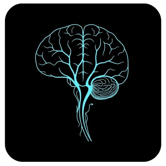
Structure of Dural Venous Sinuses
The dural venous sinuses are not typical veins but endothelial-lined channels formed between the periosteal and meningeal layers of the dura mater. Unlike veins, they lack valves, allowing for bidirectional blood flow depending on pressure gradients. These sinuses vary in size and shape, creating an interconnected system that efficiently drains blood from the brain, cranial bones, and meninges. The major sinuses include the superior sagittal sinus, inferior sagittal sinus, straight sinus, transverse sinuses, sigmoid sinuses, and the cavernous sinus. Each of these sinuses has a specific role in directing blood flow toward the internal jugular veins, facilitating efficient venous return from the cranial cavity.
Key Dural Venous Sinuses
- Superior sagittal sinusRuns along the midline on the superior aspect of the falx cerebri and drains blood from cortical veins.
- Inferior sagittal sinusLocated along the lower margin of the falx cerebri, joining the straight sinus.
- Straight sinusConnects the inferior sagittal sinus and the great cerebral vein, directing blood toward the confluence of sinuses.
- Transverse sinusesRun laterally from the confluence of sinuses, channeling blood toward the sigmoid sinuses.
- Sigmoid sinusesS-shaped channels that continue as the internal jugular veins.
- Cavernous sinusA complex sinus surrounding the pituitary gland and receiving blood from the orbit and brain.
Primary Functions of Dural Venous Sinuses
The function of dural venous sinuses extends beyond simple venous drainage. They help maintain intracranial pressure, participate in cerebrospinal fluid (CSF) absorption, and act as channels for venous blood returning to the systemic circulation. Their unique structure and location between dural layers allow for effective management of cerebral blood flow and pressure regulation.
Venous Drainage of the Brain
The most important function of dural venous sinuses is to facilitate the drainage of deoxygenated blood from the brain. They receive blood from cortical veins, deep cerebral veins, and veins of the meninges, directing it toward the internal jugular veins. This continuous drainage ensures that metabolic waste products, including carbon dioxide and other byproducts, are efficiently removed from the brain tissue, supporting optimal neuronal function.
- Drains blood from superficial and deep cerebral veins
- Transports deoxygenated blood toward the internal jugular veins
- Removes metabolic waste products from brain tissue
- Supports efficient cerebral circulation
Role in Cerebrospinal Fluid Absorption
Dural venous sinuses play a critical role in the absorption of cerebrospinal fluid. Specialized structures called arachnoid granulations or villi protrude into the sinuses, particularly the superior sagittal sinus, allowing CSF to flow from the subarachnoid space into the venous system. This process helps regulate intracranial pressure and maintains the balance between CSF production and absorption, preventing conditions such as hydrocephalus or intracranial hypertension.
- Arachnoid granulations facilitate CSF transfer into venous blood
- Helps maintain stable intracranial pressure
- Supports the balance between CSF production and drainage
- Prevents neurological complications related to fluid accumulation
Pressure Regulation and Intracranial Homeostasis
Dural venous sinuses contribute to intracranial pressure regulation by providing low-resistance pathways for venous blood and CSF drainage. Their ability to accommodate varying blood volumes helps buffer changes in intracranial pressure caused by posture, activity, or vascular events. By acting as a pressure-relieving system, the sinuses protect delicate brain tissue from damage due to venous congestion or sudden fluctuations in pressure.
- Buffers fluctuations in intracranial pressure
- Prevents venous congestion and brain tissue damage
- Supports overall cerebrovascular stability
- Maintains optimal conditions for neuronal function
Clinical Significance of Dural Venous Sinuses
Understanding the function of dural venous sinuses has important clinical implications. Thrombosis or blockage in these sinuses, known as dural venous sinus thrombosis, can lead to life-threatening conditions such as cerebral edema, increased intracranial pressure, and stroke. Imaging studies, including MRI and CT venography, are used to evaluate sinus patency and diagnose abnormalities. Additionally, knowledge of sinus anatomy is critical during neurosurgical procedures to avoid inadvertent damage and ensure safe outcomes.
Dural Venous Sinus Thrombosis
- Occurs when a blood clot forms within a dural sinus
- Can lead to headache, neurological deficits, and seizures
- Requires prompt diagnosis and anticoagulation therapy
- Understanding sinus function aids in treatment planning
Surgical and Diagnostic Considerations
- Anatomical knowledge is crucial during neurosurgery to prevent hemorrhage
- Imaging techniques assess sinus integrity and blood flow
- Evaluation of sinuses is important in conditions like trauma, tumors, or vascular malformations
Interactions with Other Structures
Dural venous sinuses interact closely with other anatomical and physiological systems. They are connected to the vertebral venous plexus, providing alternate pathways for venous drainage. Additionally, their proximity to cranial nerves and arterial structures, particularly in the cavernous sinus, highlights the importance of precise anatomical knowledge in clinical practice. These interactions ensure coordinated cerebral circulation and contribute to the brain’s overall health.
- Communicate with vertebral venous plexus for collateral drainage
- Located near cranial nerves in the cavernous sinus, affecting neurological function
- Coordinate with arterial blood flow to maintain intracranial equilibrium
The dural venous sinuses serve as essential components of the brain’s venous and cerebrospinal fluid drainage system. By facilitating efficient venous return, supporting CSF absorption, and regulating intracranial pressure, these sinuses maintain cerebral homeostasis and ensure optimal neurological function. Their complex anatomy and interconnectedness make them a critical consideration in both clinical and surgical settings. Understanding the function of dural venous sinuses highlights the remarkable ways in which the brain’s vascular system adapts to maintain balance, protect neural tissue, and sustain life.
Overall, the dural venous sinuses illustrate the intricate relationship between structure and function in human physiology. They exemplify how the body has evolved specialized mechanisms to regulate blood flow, cerebrospinal fluid dynamics, and intracranial pressure, ultimately safeguarding brain health. Recognizing their significance is essential for healthcare professionals, researchers, and students who seek to understand the complexities of cerebrovascular anatomy and its clinical implications.