Morphology Of Escherichia Coli
Escherichia coli, commonly abbreviated as E. coli, is one of the most extensively studied bacteria due to its relevance in medicine, microbiology, and biotechnology. Understanding the morphology of Escherichia coli is essential for scientists and healthcare professionals, as its structure directly impacts its behavior, reproduction, and interaction with hosts. Morphology refers to the shape, size, and structural components of the bacterium, which are crucial for identifying different strains, understanding pathogenic mechanisms, and developing treatments. The study of E. coli morphology provides insight into its adaptability and survival strategies in various environments.
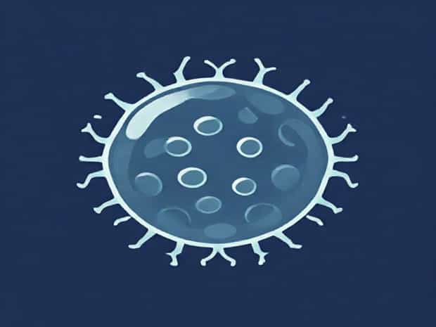
Basic Morphology of Escherichia coli
Escherichia coli is a gram-negative, rod-shaped bacterium that typically measures between 1 to 2 micrometers in length and 0.25 to 1 micrometer in diameter. Its rod-shaped or bacillus morphology allows for efficient nutrient absorption and rapid replication under favorable conditions. The cell wall is thin but structurally important, providing protection while allowing flexibility. The gram-negative nature of E. coli means that it has a complex outer membrane containing lipopolysaccharides, which play a key role in immune system interactions and pathogenicity. These morphological features are consistent across most E. coli strains, although slight variations may exist among pathogenic and non-pathogenic types.
Cell Structure Components
- Cell WallComposed mainly of peptidoglycan, it provides shape and structural integrity.
- Outer MembraneContains lipopolysaccharides (LPS) and proteins that protect against toxins and antibiotics.
- Plasma MembraneLocated beneath the cell wall, it regulates nutrient transport and metabolic processes.
- CytoplasmHouses ribosomes, enzymes, and other molecules required for cellular function.
- NucleoidThe central region containing the bacterial DNA, not enclosed by a membrane.
- FlagellaLong, whip-like structures that enable motility and chemotaxis.
- Pili or FimbriaeHair-like appendages used for adhesion to surfaces and DNA transfer during conjugation.
Flagella and Motility
One of the most distinctive features of many E. coli strains is the presence of flagella, which are long, spiral filaments extending from the cell surface. These structures are composed of the protein flagellin and are anchored in the cell membrane. Flagella enable motility through a rotary motion, allowing the bacterium to move toward favorable conditions and away from harmful substances a process known as chemotaxis. The number and arrangement of flagella vary among strains, with some possessing peritrichous flagella (distributed around the cell) and others having polar flagella (located at one or both ends). Motility is crucial for colonization and infection, especially in pathogenic strains.
Pili and Adhesion
Pili, also called fimbriae, are smaller hair-like structures that cover the surface of E. coli cells. They are primarily involved in adhesion to host tissues or surfaces, which is a critical first step in establishing infection. Some pili, known as sex pili, play a role in bacterial conjugation, allowing for the transfer of genetic material between cells. This horizontal gene transfer contributes to the spread of antibiotic resistance and other adaptive traits. Morphologically, pili are thinner and shorter than flagella, but their abundance and organization make them highly effective for surface attachment.
Gram-Negative Cell Wall and Outer Membrane
The gram-negative cell wall of Escherichia coli is a key morphological feature that influences its staining properties and susceptibility to antibiotics. Unlike gram-positive bacteria, which have thick peptidoglycan layers, E. coli has a relatively thin peptidoglycan layer sandwiched between the plasma membrane and an outer membrane. The outer membrane contains lipopolysaccharides (LPS), which function as endotoxins in pathogenic strains and trigger immune responses in host organisms. This structural complexity enhances protection against chemical attacks, detergents, and certain antibiotics, making E. coli a resilient microorganism in diverse environments.
Capsule and Surface Structures
Some E. coli strains produce a capsule, a polysaccharide layer that surrounds the cell wall. This capsule provides additional protection against desiccation, phagocytosis by immune cells, and other environmental stresses. Morphologically, the capsule can vary in thickness and composition among strains, influencing virulence and immune evasion. Additionally, surface antigens, such as O-antigens and H-antigens, are important for serotyping and distinguishing between pathogenic and non-pathogenic strains. These structural components demonstrate the diversity and adaptability of E. coli morphology.
Variations in E. coli Morphology
While the basic rod shape and gram-negative structure are consistent, E. coli exhibits morphological variations depending on environmental conditions, growth phase, and genetic differences. For example, some strains may form filamentous shapes under stress or in response to certain antibiotics. Pathogenic strains often display enhanced pili and flagella development to improve adhesion and motility. Biofilm formation, another morphological adaptation, involves clusters of cells embedded in a self-produced extracellular matrix. This structure provides protection against antibiotics and immune responses, showcasing the dynamic nature of E. coli morphology in response to its environment.
Microscopic Observation Techniques
- Light microscopy Useful for observing cell shape and general arrangements.
- Gram staining Distinguishes E. coli as gram-negative based on cell wall composition.
- Electron microscopy Provides detailed images of flagella, pili, and surface structures.
- Fluorescent microscopy Highlights specific cellular components or genetic markers.
- Atomic force microscopy Examines surface texture and mechanical properties of the cell envelope.
The morphology of Escherichia coli is a critical aspect of its biology, influencing its survival, pathogenicity, and interaction with the environment. From its rod-shaped structure and gram-negative cell wall to flagella, pili, and capsule, every morphological feature contributes to its adaptability and functionality. Variations in morphology allow E. coli to respond to environmental challenges, adhere to surfaces, and evade host defenses. Understanding these structural characteristics not only aids in microbiological research but also informs clinical approaches to infections and biotechnology applications. Studying E. coli morphology provides a foundation for appreciating the complexity and versatility of this ubiquitous bacterium.